Ultrasound
Ultrasound Exams
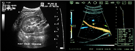
 An ultrasound exam is a safe diagnostic procedure that uses high-frequency sound waves to produce an image of many of the internal structures of the body.
Multiple studies have shown that these sound waves are harmless and may be used with complete safety, even on pregnant women, where CT or X-rays would be inappropriate. In some cases, either CT or ultrasound could be used to establish a diagnosis, but ultrasound exams are typically quicker and less expensive. Your physician will order the best kind of exam for your situation.
At BIDMC, our state-of-the-art ultrasound facilities offer:
- 18 ultrasound rooms
- Top-of-the-line equipment
- Full accreditation from the American College of Radiology and ICAVL
- A dedicated staff of technologists and physicians who will complete your study on time without unnecessary stress
The results of ultrasound exams are very much dependent on the skill of the operator. For this reason, BIDMC's technologists and doctors all have extensive ultrasound training to perform these exams.
Exams and Preparation
No preparation is necessary for these types of ultrasound exams:
- Breast
- Fetal (after fourteen weeks)
- Renal (Kidneys)
- Scrotal (Testicular)
- Thyroid (Neck)
- Amniocentesis
- Pleural aspiration
- Paracentesis
- Thoracentesis
For all other exams, please review the preparation procedures below.
An abdominal ultrasound looks at the gallbladder, bile ducts, kidneys, liver, pancreas, and spleen. It also includes a view of the aorta and retroperitoneum.

Preparation For Your Exam
Please do not eat solid food or drink anything but water for six to eight hours before the exam. Routine medications may be taken. If you are diabetic, consult with your doctor.
During the Exam
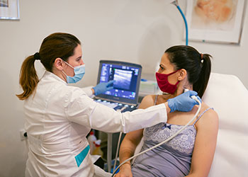
How is the exam performed? The patient lies on a table with the abdominal area exposed. The sonographer (technologist who performs the exam) will put a warm water-based gel on the skin surface. The gel helps to transmit the sound waves by excluding air. An instrument called a transducer, which is about the size of a microphone, will be moved over the skin surface by the sonographer.
How long will the exam take?
About 30 minutes.
Will it hurt?
No.
After the Exam: Getting Your Results
You may call your doctor to discuss the results.
Chorionic villus sampling (CVS) is an early prenatal test performed between the tenth and the twelfth week of pregnancy; that is ten to twelve weeks from the first day of your last menstrual period. Anyone considering CVS is required to meet with one of our genetic counselors prior to the test. They will discuss the risks and benefits of the procedure, as well as provide an opportunity to obtain a genetic family history.
Scheduling An Appointment
To schedule an appointment with a counselor or for more information, please contact the Beth Israel Deaconess Clinical Genetics Program at (617) 667-7110.
During the Exam
 The procedure, using ultrasound guidance, can be performed either vaginally or from the abdominal wall. Most are performed vaginally, but this will depend, in part, on the location of the placenta.
A thin needle or catheter is guided by ultrasound to the area where the placenta is implanting. A syringe is attached to the back of the catheter and a small sample of tissue is removed. The tissue sample obtained is about 10-25 mg in size, about the size of a very small pea. The villus tissue is of fetal origin and can be cultured by the laboratory for chromosomal analysis. When indicated, diagnostic tests for other genetic conditions may be performed.
For the procedure, we ask that you have a full bladder. This enables us to better view the pregnancy. There may be a bit of uterine pressure or cramping but most women experience little discomfort. The transabdominal procedure generally causes greater discomfort; a local anesthetic is used to numb the area.
After the Exam
 After the procedure, we recommend that you limit your physical activities that day, but many women actually return to work or travel. It is normal to have a little cramping and some scant bleeding which should subside within 48 hours. Any heavy bleeding, sharp cramps, or flu-like symptoms should be reported to your obstetrician without delay.
Results are usually available in ten days, but may require as long as two weeks. This will depend primarily on how well the cells grow in culture. It is important to note that the amount of time needed to complete the chromosomal analysis does not have anything to do with the health of the fetus.
Safety Issues
There is a one per cent risk of miscarriage associated with the CVS procedure. Also, there have been a few scientific studies that have raised concern about the incidence of certain types of birth defects in fetuses of women who have undergone this procedure. We will discuss this risk factor with you at the time of counseling.
In order to address the possibility of risks to your offspring, we recommend that you contact one of our genetic counselors to review any family history concerns and whether or not it is appropriate for you to consider prenatal diagnosis. It is important to remember that there is no one test or combination of tests that will rule out the presence of all fetal birth defects, mental retardation, or genetic disease.
Interventional ultrasound covers a broad category of diagnostic and therapeutic procedures, including biopsies, cyst aspirations, and drainage of fluid collections in the chest, abdomen and subcutaneous tissues.
Preparation For Your Exam
In general, it is preferred to avoid eating solid food and liquids other than water for 6-8 hours prior to the examination. Routine medications may be taken. If you are a diabetic, consult with your doctor regarding insulin dose.
During the Exam: How long will it take?
Interventional procedures may be preceded by diagnostic ultrasound scans. Because of the wide variety of procedures that may be performed, the time may vary from 30 minutes to 90 minutes. Certain procedures, such as biopsies of deep abdominal organs, aspiration of fluid collections in the chest, and certain other procedures may necessitate a post-procedure observation period of 1-3 hours to ensure patient well being.
Will it hurt?
Yes, there may be some pain associated with these procedures. Many can be performed with local anesthesia (similar to Novocaine used by dentists). More involved procedures may require administration of painkillers and/or anti-anxiety medications. The most involved procedures generally will involve insertion of a small intravenous line and monitoring by our Radiology nursing staff in order to administer medications intravenously. Every effort is made to minimize any discomfort during the procedure.
After the Exam
You may have some mild soreness after the procedure, but, generally, this is relatively minimal. If medications have been administered, you may feel somewhat drowsy during the recovery period and you should make arrangements for someone else to accompany you and to bring you home after the observation period, since operating machinery, such as driving, should be avoided for several hours after the procedure.
Getting Your Results
The results of the scans will be available to your physician on the day following the examination. If biopsies, cultures or other tests are performed on the material obtained during the ultrasound interventional procedure, these results may take 5-7 days to be completed. You may call your doctor to obtain results.
 Musculoskeletal ultrasound (MSK US) is a completely safe, versatile technique that uses sound waves to create an image (no radiation is involved). MSK US can be used to image any patient presenting with a complaint arising within the soft tissues of the musculoskeletal system. Any patient can have an MSK US, including those who are confined to a wheelchair or cannot have an MRI.
MSK US may be the first test to image tendon and ligament tears on the fingers and hands because it allows the doctor to see the small structures of the tendons and pulleys so well. MSK US is excellent at locating all types of foreign bodies in the soft tissues that can be difficult to detect with X-rays and MRI.
In Europe and Canada, MSK US has become the first line test to diagnose rotator cuff tears in the shoulder (rather than MRI). Studies have shown the ultrasound can be as sensitive as MRI for diagnosing rotator cuff tears and abnormalities of the biceps tendon, and it is non-invasive, much faster and cost effective to perform.
MSK US provides the additional benefit of the ability to perform dynamic imaging. During the examination, the radiologist can ask the patient to perform the maneuvers that produce the pain and document exactly what is happening. MSK US is fast and easy to do, and the image can be taken exactly where the patient says it hurts.
MSK US is also an excellent method for guiding therapeutic procedures. Under direct ultrasound guidance, physicians can directly place a needle into a joint, bursa or tendon sheath to remove fluid or inject medication exactly where it needs to go, with image documentation.
As with any test, MSK US is not perfect and does have some limitations. It is not appropriate for evaluating the labrum (the cartilage that surrounds the socket) of the hip or shoulder because the sound beam is attenuated by the overlying bony structures. MSK US should not be used to work up bone tumors and it is not as precise as MRI to characterize soft tissue tumors.
MSK US is good for the evaluation and diagnosis of abnormalities of:
- Rotator cuff, biceps tendon
- Finger and hand tendons and ligaments
- Elbow tendons and ligaments
- Carpal tunnel
- Foot and ankle tendons and ligaments
- Achilles tendon
- Plantar fascia
- Patellar and Quadriceps tendons
- Iliopsoas tendon dysfunction (snapping hip)
- Trochanteric bursitis
- Gluteus minimus and medius tendons
MSK US can also be used to evaluate a palpable soft tissue mass to determine if it is a simple cyst or warrants further workup.
MSK US should not be the first line for the evaluation of:
- Menisci of the knee, ACL, PCL
- Labrum of the hip and shoulder
MSK US should not be used to definitively characterize solid masses; patients should be referred to MRI or biopsy depending on clinical history. MSK US should not be used to evaluate bone tumors.
An obstetrical ultrasound exam looks at the uterus and ovaries, and at the fetus. The fetus is checked to be sure that its size is appropriate for its "age." The fetus is also checked to be sure the fetal anatomy is structurally normal.
Preparation For Your Exam
- You may eat regular meals prior to an obstetrical ultrasound exam.
- If you are more than fourteen weeks pregnant there is no preparation needed.
- If you are scheduled for a Biophysical Profile, please eat a meal or snack one hour before the exam.
During the Exam
 During the exam, the patient lies on a table with the abdominal area exposed. The sonographer (technologist who performs the exam) will put a warm water-based gel on the skin surface. The gel helps to transmit sound waves by excluding air. An instrument called a transducer, which is about the size of a microphone, will be moved over the skin surface by the sonographer.
How long will it take?
About 35 minutes.
Will it hurt?
No.
After the Exam: Getting Your Results
You may call your doctor to discuss the results.
Overview
A pelvic ultrasound in females looks primarily at the uterus and ovaries, but the bladder may also be visualized. In males, the pelvic ultrasound usually focuses on the bladder and the prostate gland.
Preparation For Your Exam
You may eat regular meals prior to the exam. The only special preparation is to have a full bladder at the time of the exam. Therefore, you should drink 32 ounces of water (four eight-ounce glasses, or one quart) before the exam. Start drinking it one hour before the exam, and finish drinking it half an hour before the exam. Do not urinate before the exam.
During the Exam
The patient lies on a table with the abdominal area exposed. The sonographer (technologist who performs the exam) will put a warm water-based gel on the skin surface. The gel helps to transmit sound waves by excluding air. An instrument called a transducer, which is about the size of a microphone, will be moved over the skin surface by the sonographer. When more detailed views of the uterus, ovaries or surrounding tissues are required, a special sterilized high resolution probe may be utilized by scanning through the vagina.
How long will it take?
About twenty minutes.
Will it hurt?
No.
After the Exam: Getting Your Results
You may call your doctor to discuss the results.
Transrectal ultrasound allows the radiologist to closely examine the prostate gland for abnormalities. At certain times, multiple biopsies of the prostate gland may be performed to examine for any evidence of cancer or inflammation.
Sometimes transrectal ultrasound of the prostate is used to diagnose the cause of male infertility.
Preparation For Your Exam
For the transrectal prostate exam, a "Fleet" enema should be taken about four hours before the exam. This preparation with instructions can be bought from your local pharmacy. If a biopsy is going to be performed, as it usually will be, your doctor will give you some antibiotics to take, usually Cipro. One tablet should be taken the night before the biopsy, one the morning of the biopsy, one that evening and one the following morning. Cipro is expensive, approximately $2 to $3 a tablet, but is currently the best antibiotic for this procedure.
During the Exam: How is the exam performed?
An ultrasound transducer about the size of a finger is placed into the rectum and the prostate is scanned.
How long will the prostate exam take?
A prostate exam with or without the biopsy will take about 15 to 25 minutes.
What happens in the ultrasound room?
After changing into hospital garb you will be escorted into the ultrasound exam room. You will be asked to lie on a table on your left side with your knees bent. A transducer will be carefully inserted into your rectum by the sonographer or radiologist. Pictures and measurements of the prostate will then be taken. If a biopsy is to be performed either the urologist or the radiologist will then take between six to eight biopsies. A small needle is inserted very rapidly into the prostate gland. The tissue will be sent to the pathology department for preparation.
Will it hurt?
Yes, but not very much and not as much as most people think. The prostate is not very sensitive to needle sticks. The vast majority of men tolerate the procedure very well -- typically the anxiety they experience prior to the biopsy is considerably worse than the biopsy itself.
After the Exam
You may experience some mucous or a small amount of bleeding from your rectum after the prostate exam. There may be some minor discomfort after a biopsy and some small amount of blood in the urine or rectum for up to 48 hours. Blood in the semen is also common. These are not cause for concern if the amounts are small.
Getting Your Results
The results of the prostate ultrasound will be available to your doctor on the day following your exam. Biopsy results are generally ready five to seven days after the procedure. If your urologist has not given you the results within one week, we suggest you telephone his or her office and ask for the results.
Rectal ultrasound can be used to evaluate rectal polyps and masses, in particular to evaluate the extent of involvement of the rectal wall and the perirectal tissues. This information helps the surgeon in planning the most effective surgical approach to removal of the lesion. This study can also be used in followup of patients who have had rectal tumors resected, and is also utilized to evaluate for inflammatory and infectious conditions surrounding the rectum.
Anal ultrasound is used to evaluate the anal sphincter musculature in patients with incontinence or constipation, as well as to evaluate inflammatory and infectious conditions affecting the anal canal, such as fistulas.
Preparation For Your Exam
A mini "Fleets" enema is taken just before the examination in order to provide a clean rectal wall through which to scan. No other preparation is necessary.
During the Exam
After changing into hospital garb, you will be escorted into the Ultrasound Examination Room. You will be asked to lie on a table on your left side with your knees bent. A transducer will be carefully inserted into your rectum by the sonographer or radiologist and the scans and images will then be obtained, by moving the probe forward and backward through the anal canal and rectum.
How long will the exam take?
A rectal or anal ultrasound generally takes 5-10 minutes time.
Will it hurt?
The ultrasound transducer is approximately the size of a finger. While there is some brief discomfort as the probe is inserted, most patients find the discomfort is very minimal and of brief duration. For rectal scans, once the probe is past the anal sphincter, a water-filled balloon is inflated to optimize the image quality, but this does not cause any pain or discomfort. For anal ultrasound, the probe remains in the anal canal as the images are obtained, and this is slightly uncomfortable, but this study is generally completed within 2-3 minutes.
After the Exam: Getting Your Results
The results of the transrectal or transanal ultrasound will be available to your doctor on the day following your exam. You may call your doctor to discuss the results.
High resolution small parts ultrasound is most frequently utilized to evaluate for possible abnormalities in the thyroid and parathyroid glands, in the scrotum and testis, in the breast, and occasionally at other superficial sites of possible abnormality. The study not only allows visualization and characterization of abnormalities, but can also be utilized to guide fine needle aspirations and biopsies of possible abnormalities.
Preparation For Your Exam
No special preparation is necessary.
During the Exam: How is the exam performed?
The patient lies on a scanning table with the area to be examined exposed. The sonographer (technologist who performs the exam) will put a warm water-based gel on the skin surface which helps to transmit sound waves by excluding air. An instrument called a transducer, which is about the size of a microphone, will be moved over the skin surface by the sonographer to obtain the diagnostic images.
How long will the exam take?
These exams typically take approximately 15-20 minutes time.
Will it hurt?
No.
After the Exam: Getting Your Results
The results of the scan will be available to your doctor on the day following your examination. If biopsies are performed, the results are generally available 5-7 days after the procedure.
Liver, kidney and pancreatic transplant evaluation is provided in order to evaluate the transplanted organ for any signs of rejection or other abnormalities.
During the Exam: How is the exam performed?
The patient lies on a table with the abdominal area exposed. The sonographer (technologist who performs the exam) will put a warm water-based gel on the skin surface, which helps to transmit the sound waves by excluding air. An instrument called a transducer will be passed over the abdominal surface in order to obtain the images.
How long will the exam take?
A typical examination may take approximately 30 minutes time.
Will it hurt?
No.
After the Exam: Getting Your Results
Results of the ultrasound study will be available to your doctor on the day following the exam. You may call your doctor to discuss the results.
A vascular ultrasound exam looks at the blood vessels to see whether there are any areas of dilatation, narrowing, or blockage. The vessels most frequently looked at are in the neck, arms, and legs, including both arteries and/or veins, as well as assessment of bypass grafts and AV fistulas for hemodialysis.
Preparation For Your Exam
No special preparation is necessary.
During the Exam: How is the exam performed?
The patient lies on a table with the abdominal area exposed. The sonographer (technologist who performs the exam) will put a warm water-based gel on the skin surface. The gel helps to transmit sound waves by excluding air. An instrument called a transducer, which is about the size of a microphone, will be moved over the skin surface by the sonographer.
How long will the exam take?
Probably about a half hour, depending on the area to be studied. In some cases the exam may last up to one and a half hours.
Will it hurt?
No.
After the Exam: Getting Your Results
You may call your doctor to discuss the results.
