Robotic Pyeloplasty
Understanding Robotic Pyeloplasty
Blockage of urine outflow from the kidney is known as a ureteropelvic junction (UPJ) obstruction; it typically is caused by congenital scarring or a blood vessel pressing on the ureter. This can result in pain, infection, kidney stones and loss of kidney function.
Pyeloplasty, or surgical removal of the UPJ obstruction, returns the
outflow of urine to normal. Traditionally, the procedure has been performed
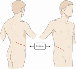
as an open surgery through an 8-12 inch incision in the flank. (see figure
1a and 1b) A minimally invasive alternative, robotic pyeloplasty has been
demonstrated to have success equal to that of traditional open surgery but
with added benefits: less postoperative pain, a shorter hospital stay,
earlier return to normal activity and a better cosmetic result.1-2

Figure (right)
Traditional open kidney surgery is performed through an 8-12 inch incision
extending from the ribs towards the abdomen. A portion of one of the ribs
is usually removed as part of the surgery.
The procedure
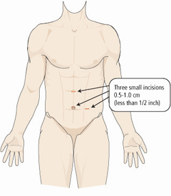
Robotic pyeloplasty requires a general anesthetic. First, 3 tiny "keyhole" incisions, each 1cm, are made in the patient's abdomen. A laparoscopic camera and thin, specialized instruments are placed through these incisions. The area of obstruction then is isolated and either removed or repaired using laparoscopic suturing techniques. A ureteral stent is left in place for 3 weeks following the surgery and removed in the urologist's office. (see figure 2)
Advantages over open surgery
- Less pain
- Shorter hospital stay
- Quicker recovery
- Better cosmetic result
Results
Cure rate
Patients should expect a greater than 95% success rate,1-2 equal to that of
the open procedure if performed by an experienced surgeon such as those at
BIDMC.

Figure (right)
Robotic-assisted pyeloplasty is performed through 3 small incisions in the
abdomen.
Blood loss
Typical blood loss is 100cc. Blood transfusions are extremely rare.
What to Expect after Surgery
Hospital stay
The typical stay is 2 days.
Diet
Patients resume normal eating as they recover in the first day after surgery or soon thereafter.
Postoperative pain
Pain is managed immediately with an intravenous patient controlled analgesia pump, which is removed and replaced by pills the day after surgery. Upon discharge, pain pills are given for several days, after which over-the-counter acetaminophen or ibuprofen are usually all that is needed.
Urinary catheter
A urinary catheter is left in place for one or two days following the surgery and then removed.
Abdominal drain
A small drain placed in the area of the kidney is usually removed the second day after surgery.
Recovery
Time to complete recovery is typically 3-4 weeks compared with 8 weeks for
open pyeloplasty.

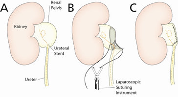
Figure (right)
A: kidney, renal pelvis, and ureter shown prior to pyeloplasty. Note the
distended renal pelvis as a result of urine obstruction and the ureteral
stent placed just prior to the procedure.
B: the obstructed portion of the ureter has been removed and the ureter is
sutured back to the renal pelvis using laparoscopic suturing techniques.
C: This is the kidney and ureter following the procedure. A smooth funnel
shape is recreated to allow urine to flow freely from the kidney. The
ureteral stent is removed in 3 weeks.
Follow-up
Patients will follow-up with their surgeon 1 month after the operation for a routine visit, then at 3, 12 and 24 months with nuclear renal imaging and ultrasounds of the kidney. Appointments can easily be conducted over the phone for patients living outside the Boston area, including international patients.
References
1. Moon DA El-Shazley MA, Chang CM, Gianduzzo TR, Eden CG. "Laparoscopic pyeloplasty: evolution of a new gold standard." Urology, May 2006: 67: 932-6
2. Romero FR, Wagner AA, Trapp C, Permpongkosol S, Link RE, Kavoussi LR. "Tansmesenteric laparoscopic pyeloplasty." The Journal of Urology, 2006 in press
