About Our Imaging Center
Our patients’ comfort and providing state-of-the-art imaging care are our top priorities.
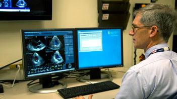
The noninvasive cardiac imaging and testing specialists in the CVI provide a range of cardiovascular imaging services for individuals with suspected or existing heart and vascular conditions.
Cardiovascular imaging is an integral part of diagnosing a variety of cardiovascular conditions, and is often used to monitor the effectiveness of treatment. Each year, BIDMC specialists perform more than 70,000 electrocardiograms, 15,000 echocardiograms, 5,000 stress tests, 800 cardiac magnetic resonance imaging studies and 1,800 radionuclide studies.
Our specialists work closely with other cardiovascular medicine colleagues, primary care physicians, radiologists and anesthesiologists. This collaboration among multiple medical disciplines keeps the lines of communication open and enhances quality of care for patients and families.
Types of Imaging
Cardiac MRIAppointments: 617-667-8555
Cardiovascular MRI (CMR) is specialized MRI of the heart and vascular system, requiring particular equipment and expertise.
The CVI's Cardiac Magnetic Resonance Imaging (CMR) Center is a joint effort of the Division of Cardiovascular Medicine and Department of Radiology. Our Cardiac Magnetic Resonance Center is a world leader in the development and clinical use of:
- CMR for coronary artery imaging
- Using CMR to assess cardiomyopathies, pericardial disease and congenital heart disease
The center features a state-of-the-art Tesla Siemens (MAGNETOM Vida) cardiac magnetic resonance imaging system, which provides improved patient comfort and enhanced image quality.
We are committed to the education and training of researchers and clinicians with funding provided by grants from the National Institutes of Health, National Heart, Lung and Blood Institute, American Heart Association, industry, private foundations and individual contributions.
Clinical Physiology Stress TestingAn exercise stress test helps your doctor to determine how well your heart functions during physical activity; specifically, whether it is getting enough blood and oxygen when it is working hard. During the test, you will walk on a treadmill while the doctor monitors your heart function.
The CVI's Cardiovascular Clinical Physiology Stress Testing Laboratory has a national and international reputation as a key center for the use of stress testing as an outcome measurement. Our multidisciplinary staff is comprised of physicians, physiologists and technologists.
Patients are referred to the lab to determine the cause, risk assessment and outlook associated with various forms of heart disease, such as ischemic syndrome, congestive heart failure, arrhythmia and valvular heart disease.
Physicians typically order stress tests for patients with:
- Chest pain
- Unexplained shortness of breath
- Unexplained arm, neck, back or jaw discomfort
- Fatigue or weakness
- Palpitations
- Dizziness
- Fainting episodes
- Valvular heart disease
- Heart failure
Types of Stress Tests
The lab features state-of-the-art equipment to perform a range of treadmill, bicycle and pharmacologic tests, including:
- Exercise testing without imaging
- Radionuclear perfusion (blood flow within the heart)
- Echocardiography: wall motion or gradient to assess valve function or blood flow to the heart
- Pharmacologic or pacing stress tests for patients who are unable to perform functional stress tests
- Cardiopulmonary testing: metabolic (oxygen consumption) and oxygen saturation testing in conjunction with select stress testing to determine cardiac function
- T-wave alternan, a computer-based program that allows the clinician to detect patients at risk for sudden cardiac death syndrome
Cardiovascular CTAppointments: 617-667-2515
Cardiovascular Computed Tomography (CCT) is a collaborative effort between the Division of Cardiovascular Medicine and the Department of Radiology, which pioneered this non-invasive method to evaluate coronary artery disease. The laboratory continues to have the most up-to-date technology.
CCT is a painless diagnostic method in which a dye is injected into the arm vein. As this dye circulates through the body, the image of the heart is captured in just a few beats. A computer then displays the information three-dimensionally to help your radiologist and physician determine if you have coronary artery disease or other abnormalities of the heart and vascular system.
EchocardiographyAn echocardiogram is a medical test that produces detailed images of your beating heart. Using ultrasound technology, a transthoracic echocardiogram allows healthcare providers to see the walls, chambers and valves within your heart, as well as blood flow within your heart. The test is not invasive and does not use any radiation or require any sedation.
The CVI's Echocardiography Laboratory has been fully accredited by the Intersocietal Accreditation Commission of Echocardiography Laboratories (ICAEL) continuously for 10 years, and all our our interpreting physicians have passed the American Board of Echocardiography Special Competency in Echocardiography examination.
Our lab features state-of-the-art imaging systems with real-time electronic report generation.
Procedures
- Transthoracic (surface) echocardiography
- Transesophageal echocardiography
- Stress (physiologic and pharmacologic) echocardiography
- Contrast echocardiography
You may be referred to the laboratory if your doctor has diagnosed or suspects one of the following diseases and conditions:
- Valvular heart disease
- Ventricular dysfunction
- Congenital heart disease
- Cardiomyopathy
- Congestive heart failure
- Pericardial disease
- Cardiac source of embolism (clot)
- Coronary artery disease
ElectrocardiographyElectrocardiograms (ECGs or EKGs) are a record of the electrical activity of the heart. This noninvasive, painless test is an important diagnostic tool for clinicians, a means to monitor the results of therapy, and a tool to detect changes in both acute and chronic heart conditions.
The CVI's Electrocardiography Lab serves as an important teaching and training resource for the development of expertise in ECG interpretation by cardiology trainees as well as Harvard Medical students on the cardiology elective rotations.
Nuclear CardiologyAppointments: 617-667-2071
A radionuclide cardiac perfusion study is a minimally invasive tool that uses small amounts of radioactive materials to create images of the heart’s blood flow. Radionuclide studies allow physicians to evaluate the heart’s pumping action and the extent and severity of coronary artery disease, including heart attacks.
Nuclear medicine exams use the opposite approach of typical X-rays and CT scans: a radioactive material is introduced into the patient's body (usually by injection), and is then detected by a machine called a gamma camera.
Radionuclide studies also help doctors assess chest pain (angina) and evaluate the success of bypass surgery or stent placement, among other things.
The CVI’s Nuclear Cardiology Laboratory is a collaborative effort between the Division of Cardiovascular Medicine and the Division of Nuclear Medicine Division in BIDMC’s Department of Radiology. Our laboratory provides diagnostic services for outpatients and inpatients, including,cardiac perfusion exams using rapid heart scans.
The Nuclear Medicine Division at BIDMC is fully accredited by the American College of Radiology (ACR).
Obtaining Your Images
To receive a copy of exam images, please follow these steps:
- Email us or call 617-632-7360. Please provide your name and other identifying information along with the date of the study being requested.
- Fill out and sign a Medical Images Release form.
- Email the completed form to us or fax it to 617-632-7620.
- We can send the images electronically to you or by mail. If you would like us to send a copy of your study to a physician outside the BIDMC, we will need a signed release from you along with the study you would like sent and the receiving doctor's name and email or address. You can send us that signed release form to the email or fax number listed above.
More on Non-Invasive Cardiac Imaging
Accredited Facility for Echocardiography
The CVI's Echocardiography Laboratory has been fully accredited by the Intersocietal Accreditation Commission of Echocardiography Laboratories (ICAEL) continuously for 10 years.