Diagnosing Brain Aneurysm
Advanced Imaging Technology Aids Diagnosis
The BIDMC Advantage
 The Brain Aneurysm Institute at Beth Israel Deaconess Medical Center uses advanced imaging technology — invasive and noninvasive — to help doctors make the best diagnosis and to guide treatment. These highly sophisticated images can pinpoint the location, type, size and other particulars about the aneurysm to determine the risks and benefits of therapy. Doctors also use these same tests to zero in on other complex brain and spine vascular disorders.
Both CT and MRI scans at BIDMC feature perfusion imaging, an advanced capability to assess abnormal blood vessel formation, the rate of blood flow, blood flow abnormalities, changes caused by disease, or changes in response to treatment. We are leaders in developing and advancing MRI perfusion imaging, and our neuroradiologists are highly skilled at interpreting the results.
Because our noninvasive imaging is at such a high level at BIDMC, we often can spare patients from undergoing arteriography and its associated risks for diagnostic purposes only. We glean the diagnostic information we need in most cases from CT or MRI scans. We reserve invasive catheter angiography for patients who are undergoing an endovascular treatment at the same time, or when noninvasive tests show the need for specific information only available from the catheter technique.
Imaging Tests Used for Diagnosis
CT Scan (Computed Tomography)
An X-ray image of the head, processed by a computer into two- and three-dimensional images of the skull and brain. A CT scan can show the presence of an aneurysm and, if the aneurysm has burst, detects blood that has leaked into the brain.
CT Angiography
Combines a CT scan with contrast dye injected into the bloodstream. CT(A) is considered noninvasive because it involves only an injection into a vein. Compared to a standard CT scan, this test produces clear and detailed images of the brain arteries. The images help doctors detect and analyze aneurysms and other abnormalities of the blood vessels.
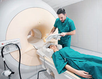
MRI (Magnetic Resonance Imaging)
 MRI uses a powerful magnetic field (not radiation as do X-rays and CT scans) to create brain images. These can be valuable in determining the effects on the brain of blood vessel abnormalities and the anatomy of aneurysms and other vascular lesions.
MRI Angiography
This type of imaging uses MRI to visualize the brain arteries and veins. Depending on the information needed, this may or may not require an intravenous injection. MRA is complementary to CT(A) images for displaying two-, three- and four-dimensional images of blood vessels.
We provide magnetic resonance imaging (and CT as well) 24 hours a day, 7 days a week, for any patient who needs these studies.
We also have sophisticated CT-MRI image fusion technology. This process allows doctors to collect and combine patient data from two different studies — CT and MRI — and fuse them together into one three-dimensional rendering for improved clinical accuracy.
Cerebral or Spinal Angiogram
Also called an arteriogram or catheter angiogram, this invasive test requires numbing anesthesia and a small incision in the groin. Doctors insert a catheter (a thin, flexible tube) through arteries from the groin to the neck. Contrast dye injected into the bloodstream highlights any abnormalities: an aneurysm, vascular malformation, or an obstruction or narrowing in a blood vessel in the neck, head or brain. This test can help diagnose stroke, unruptured and ruptured aneurysms, and other brain and spine vascular abnormalities. Angiography is the first step in endovascular treatment of these abnormalities, and is often performed before radiation or surgical therapy.
Lumbar Puncture/Spinal Fluid Analysis
A test doctors may order when they suspect a ruptured aneurysm. Using a needle, doctors remove a small amount of fluid from the space between the spinal cord and its protective covering. Blood in the fluid would indicate bleeding or brain hemorrhage, and a likely ruptured aneurysm.
